Cytoplasm Segmentation in High-Resolution Microscopy with Deep Block

DeepBlock.net is a state-of-the-art deep learning platform that has been successful in overcoming challenges in analyzing microscope images of cytoplasm.
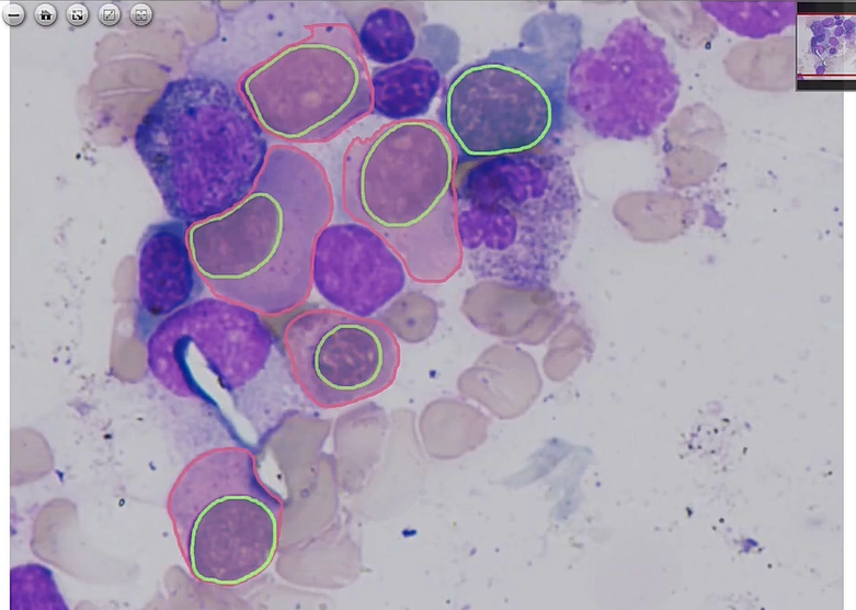
Check our latest machine learning project for cancer cell segmentation for high resolution microscopy.
https://app.deepblock.net/deepblock/store/project/plilkergjlqxlprn8
Definition of Cytoplasm
The cytoplasm is a gel-like substance that fills the cell and surrounds the organelles. It is a vital component of the cell and plays a crucial role in various cellular processes. Cytoplasm consists of water, proteins, lipids, carbohydrates, ions, and other molecules.
The main function of cytoplasm is to provide a medium for the organelles to carry out their functions. It also helps in the transport of molecules within the cell and provides structural support.
Understanding the definition of cytoplasm is important to comprehend its significance in cell biology and the study of cellular processes.
The Importance of Investigating Cytoplasmic Morphology
Investigating cytoplasmic morphology is crucial to gain insights into the structure and function of cells. The morphology of the cytoplasm can vary depending on the cell type, stage of development, and cellular processes occurring within the cell.
Studying cytoplasmic morphology can provide valuable information about cell health, cellular organization, and the presence of abnormalities or diseases. It can also help in understanding the mechanisms underlying cellular processes such as cell division, movement, and signaling.
By investigating cytoplasmic morphology, researchers can unravel the complexities of cellular structures and functions, leading to advancements in various fields such as medicine, bioengineering, and biotechnology.
Microscopic Examination of Cytoplasm
Microscopic examination is a powerful tool in studying the structures within the cytoplasm. With the use of advanced microscopes, researchers can visualize the intricate details of the cytoplasmic components.
Microscopy techniques such as light microscopy, electron microscopy, and fluorescence microscopy enable researchers to observe different aspects of cytoplasmic structures. These techniques provide information about the size, shape, distribution, and dynamics of organelles, cytoskeletal elements, and other cellular components.
By examining the cytoplasm at a microscopic level, researchers can gain a deeper understanding of cellular processes and the interactions between different cellular components.
Advantages of Implementing Automated Cytoplasm Segmentation
Automated cytoplasm segmentation is a technique that utilizes image processing algorithms to identify and separate cytoplasmic regions from microscopic images.
Implementing automated cytoplasm segmentation offers several advantages. It reduces the time and effort required for manual segmentation, enables the analysis of a large number of cells in a high-throughput manner, and provides quantitative measurements of cytoplasmic features.
Automated cytoplasm segmentation also helps in standardizing the analysis process and reducing potential bias introduced by manual segmentation. It allows researchers to focus on the analysis and interpretation of cytoplasmic data rather than spending significant time on segmentation tasks.
Overall, implementing automated cytoplasm segmentation enhances the efficiency and accuracy of cytoplasmic analysis, facilitating advancements in cell biology research.
Difficulties in Analyzing High-Resolution Microscope Images of Cytoplasm
Analyzing high-resolution microscope images of cytoplasm can pose several challenges. The size of high-resolution microscopic images, the file manipulation of microscopic images, and computation and deep learning techniques can make automation difficult.
Segmenting cytoplasmic regions accurately from high-resolution images requires advanced image processing algorithms and computational techniques. The processing of files for rendering, inference interface, and the specially tailored machine learning model and algorithm for microscopy present considerable challenges.
Despite these challenges, advancements in image analysis algorithms and computational resources have significantly improved the analysis of high-resolution microscope images of cytoplasm.
The Success of DeepBlock.net in Overcoming Challenges in Analyzing Microscope Images of Cytoplasm
DeepBlock.net is a state-of-the-art deep learning platform that has been successful in overcoming challenges in analyzing microscope images of cytoplasm.
By utilizing deep neural networks, DeepBlock.net can accurately segment cytoplasmic regions from high-resolution images. It can handle complex cytoplasmic structures, variations in staining and imaging techniques, and the presence of noise and artifacts.
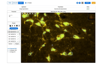
DeepBlock.net also offers annotation tools, enabling researchers to customize their own machine learning model for microscopic images. It provides a user-friendly interface and efficient computational capabilities, making the analysis of large image datasets more accessible.
The success of DeepBlock.net in overcoming challenges in analyzing microscope images of cytoplasm opens up new possibilities for cell biology research and facilitates the discovery of novel cellular structures and functions.


