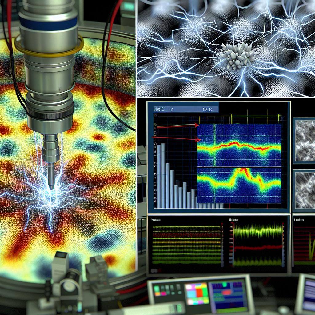
Understanding Electron Beam-Induced Charging in Microscopy

Explore how electron beam-induced charging can impact microscopy analyses and discover strategies to mitigate these effects for clearer, more accurate observations.
The Basics of Electron Beam-Induced Charging
Electron beam-induced charging is a phenomenon that occurs when a sample is exposed to an electron beam in microscopy. When the electron beam strikes the sample, it can cause the sample to become electrically charged.
This charging effect is a result of the interaction between the high-energy electrons in the beam and the atoms or molecules in the sample. The electron beam transfers energy to the sample, causing the atoms or molecules to gain or lose electrons and become charged.
The charging effect can be influenced by various factors, including the composition and structure of the sample, the intensity and duration of the electron beam, and the environmental conditions in the microscope.
Understanding the basics of electron beam-induced charging is crucial for performing accurate microscopy analyses and obtaining reliable results.
How Charging Affects Microscopy Images
Charging in microscopy can have significant effects on the resulting images. When a sample becomes charged, it can distort the electron beam, leading to changes in the image formation process.
One of the primary effects of charging is the creation of electrostatic forces that can cause the sample to attract or repel the electron beam. This can result in image distortion, blurring, or even complete loss of contrast.
Charging can also lead to the accumulation of charge at specific areas of the sample, causing localized variations in the image intensity. These variations can obscure details and make it difficult to accurately interpret the microscopy data.
Additionally, charging can induce changes in the surface potential of the sample, affecting the secondary electron emission process. This can further impact the image formation and result in changes in the overall image quality.
It is essential to understand the effects of charging on microscopy images to avoid misinterpretation of the data and to develop strategies for minimizing these effects.
Techniques to Mitigate Charging in Electron Microscopy
To mitigate charging effects in electron microscopy, various techniques can be employed. These techniques aim to reduce or eliminate the charging-induced artifacts and improve the image quality and accuracy of the observations.
One common approach is the use of conductive coatings or materials on the sample. These coatings provide a path for the dissipation of the charge, preventing its accumulation and reducing the impact on the image formation process.
Another technique is the use of anti-static devices, such as ion guns or neutralizers, which neutralize the charge accumulated on the sample surface. These devices can help minimize the effects of charging and improve the overall image quality.
Adjusting the beam energy and current can also help mitigate charging effects. Lowering the beam energy or reducing the beam current can minimize the transfer of energy to the sample, reducing the charging effect.
Optimizing the environmental conditions in the microscope, such as controlling the humidity or using a vacuum, can also help mitigate charging effects. These conditions can influence the charging behavior of the sample and minimize its impact on the image formation process.
By employing these techniques and implementing appropriate mitigation strategies, researchers can overcome the challenges posed by charging in electron microscopy and obtain clearer and more accurate observations.
Case Studies: Overcoming Charging Challenges
Several case studies have demonstrated successful strategies for overcoming charging challenges in electron microscopy.
For example, researchers have used conductive coatings on non-conductive samples to mitigate charging effects in SEM imaging. The conductive coating provides a path for charge dissipation, reducing the accumulation of charge and minimizing image distortions.
In another case study, researchers utilized an anti-static device in TEM imaging to neutralize the charge accumulated on the sample surface. This helped improve the image quality and obtain clearer observations.
These case studies highlight the importance of understanding and addressing charging challenges in electron microscopy and provide valuable insights into effective mitigation strategies.


