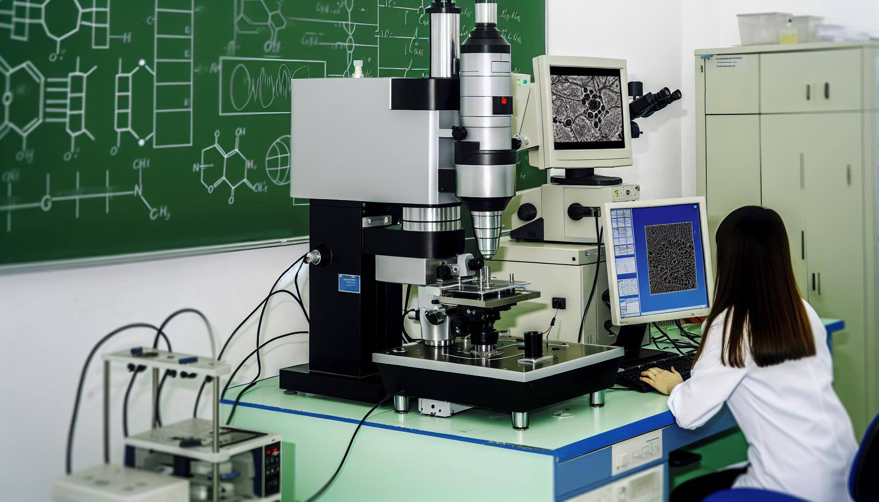
Mastering Fixation Techniques for Electron Microscopy

Unlock the secrets of perfect electron microscopy samples with advanced fixation techniques. Discover how these methods can dramatically improve your microscopy results.
Understanding the Role of Fixation in Electron Microscopy
Fixation is a crucial step in electron microscopy sample preparation. It involves preserving the structure and morphology of the sample, allowing for detailed analysis under the electron microscope. By immobilizing the sample and preventing any changes or degradation during the imaging process, fixation ensures accurate and reliable results.
Fixation also helps to maintain the sample's ultrastructure, preserving cellular components and organelles in their natural state. This is particularly important for biological samples, where the delicate structures can be easily damaged or altered.
Different fixation techniques can have varying effects on the sample, so it is essential to understand their roles and choose the most suitable method for your specific research needs.
Comparing Chemical and Physical Fixation Methods
Chemical fixation and physical fixation are two main categories of fixation methods used in electron microscopy. Each method has its advantages and limitations, and choosing the right one depends on the nature of the sample and the research objectives.
Chemical fixation involves treating the sample with fixatives such as aldehydes, which crosslink proteins and preserve the cellular structure. This method is widely used for biological samples and allows for excellent preservation of cell morphology. However, it can introduce artifacts and may alter the chemical composition of the sample.
On the other hand, physical fixation techniques involve freezing or cryopreserving the sample to immobilize its structure. This method is particularly useful for samples that require preservation of dynamic processes or delicate ultrastructures. It avoids the chemical reactions associated with chemical fixation but may introduce ice crystal artifacts.
By comparing the advantages and limitations of chemical and physical fixation methods, researchers can make informed decisions to achieve the best results for their specific samples.
Step-by-Step Guide to Chemical Fixation for Biological Samples
Chemical fixation is a widely used method for preserving the cellular structure of biological samples in electron microscopy. Follow these step-by-step instructions to achieve optimal results:
1. Choose the appropriate fixative: Select a fixative that is suitable for your specific sample and research objectives. Common fixatives include glutaraldehyde and paraformaldehyde.
2. Prepare the fixative solution: Prepare a fresh fixative solution according to the manufacturer's instructions or established protocols. Ensure the solution is properly buffered to maintain the sample's pH.
3. Pre-treat the sample: Depending on the sample type, it may require pre-treatment steps such as washing or dehydration. Follow the recommended protocols to prepare the sample for fixation.
4. Fixation process: Immerse the sample in the fixative solution and allow it to penetrate the cells. The duration of fixation varies depending on the sample size and type. It is crucial to follow the recommended fixation time to avoid over-fixation or under-fixation.
5. Post-fixation steps: After fixation, rinse the sample with buffer solution to remove excess fixative. This step helps to minimize artifacts and improve the quality of the final imaging results.
By following this step-by-step guide, researchers can achieve consistent and reliable results with chemical fixation techniques.
Innovations and Best Practices in Physical Fixation
Physical fixation techniques have seen significant advancements in recent years, offering researchers new possibilities for preserving delicate samples in electron microscopy. These innovations aim to overcome the limitations of traditional physical fixation methods and improve the quality of imaging results.
One such innovation is the use of cryo-fixation, which involves rapidly freezing the sample to preserve its structure. Cryo-fixation enables the study of dynamic processes and fragile samples without the artifacts introduced by chemical fixatives. It allows for high-resolution imaging of biological samples and provides valuable insights into cellular processes.
Another best practice in physical fixation is the use of cryo-embedding techniques, where the sample is embedded in a vitrified ice matrix. This technique eliminates the formation of ice crystals, which can distort the sample's ultrastructure. Cryo-embedding allows for the preservation of cellular morphology and provides a more accurate representation of the sample's natural state.
By staying updated with the latest innovations and adopting best practices in physical fixation, researchers can enhance the quality and reliability of their electron microscopy results.
Troubleshooting Common Fixation Issues and Tips for Success
Fixation in electron microscopy can sometimes pose challenges, leading to suboptimal results. Understanding common fixation issues and implementing troubleshooting strategies can help researchers achieve success in their experiments.
One common issue is over-fixation, where the sample becomes excessively crosslinked and rigid. This can result in loss of fine structural details and poor contrast. To avoid over-fixation, it is important to follow the recommended fixation times and concentrations.
Under-fixation is another challenge that can lead to poor preservation of the sample's ultrastructure. It is crucial to ensure that the fixative adequately penetrates the sample and that the recommended fixation time is followed.
Artifacts, such as precipitates or distortions, can also occur during fixation. These artifacts can interfere with the interpretation of imaging results. To minimize artifacts, researchers should carefully choose the appropriate fixative and optimize the fixation conditions for their specific samples.
Proper sample handling and storage are also essential for successful fixation. Samples should be handled gently to avoid damage, and they should be stored in appropriate conditions to maintain their integrity.
By troubleshooting common fixation issues and implementing best practices, researchers can overcome challenges and achieve reliable and high-quality results in electron microscopy.


