Explore the world of microtomes in microscopy, from ultramicrotomes to laser microtomes.
Understanding Microtomes: Purpose and Function
Microtomes are laboratory instruments used to accurately cut thin sections of samples for microscopic examination. They are commonly used in histology, pathology, and other fields that require detailed analysis of tissue samples.
The main purpose of a microtome is to produce thin sections of samples that can be mounted on microscope slides under a microscope. This allows researchers to study the structural details of the sample.
There are various types of microtomes available, each with its own advantages and applications. Understanding the purpose and function of different microtomes is essential for selecting the right instrument for specific laboratory needs.
Ultramicrotomes: Advanced Sectioning for Electron Microscopy
Ultramicrotomes are a type of microtome that are specifically designed for advanced sectioning in electron microscopy. These microtomes are capable of cutting ultra-thin sections of tissue samples, typically ranging from 30 to 70 nanometers in thickness.
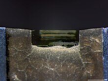
One of the key features of ultramicrotomes is their ability to use an ultramicrotome diamond knife, which is an extremely sharp blade that can cut tissue samples with high precision. This allows researchers to obtain thin sections of samples that are suitable for electron microscopy imaging.
Ultramicrotomes also often come equipped with a cryo-chamber, which allows for sectioning of frozen tissue samples. This is particularly useful for preserving the integrity of delicate tissues and structures that may be damaged during the traditional embedding and sectioning process.
In summary, ultramicrotomes are essential tools in electron microscopy, enabling researchers to obtain ultra-thin sections of samples with high precision and preserving the integrity of delicate structures.
Rotary Microtomes: Precision in Histological Techniques
Rotary microtomes are widely used in histological techniques for the preparation of tissue sections. These microtomes utilize a rotating knife to make thin and consistent sections of tissue samples.
One of the key advantages of rotary microtomes is their ability to produce sections with a high degree of precision and consistency. This is crucial for histological analysis, as it allows for accurate comparisons and measurements of tissue structures.
Rotary microtomes also offer adjustable section thickness, making them versatile instruments that can be used for a wide range of applications. The thickness can be easily adjusted using a micrometer mechanism, allowing researchers to obtain sections of varying thickness as required.
In addition to their precision and versatility, rotary microtomes are also known for their durability and ease of use. They are commonly used in research laboratories, pathology departments, and educational institutions for histological studies and research.
Cryostat Microtomes: Enabling Frozen Section Analysis
Cryostat microtomes are specialized microtomes that are designed to cut frozen tissue samples. These microtomes are commonly used in research and diagnostic laboratories for frozen section analysis.
One of the key advantages of cryostat microtomes is their ability to preserve the integrity of delicate tissue structures during the sectioning process. By cutting the tissue while it is frozen, cryostat microtomes minimize the risk of tissue damage and provide high-quality sections for analysis.
Cryostat microtomes are equipped with a temperature-controlled chamber that maintains the tissue sample at a low temperature, typically around -20°C to -30°C. This freezing temperature allows the tissue to harden, making it easier to obtain thin sections without distortion or artifacts.
The sections obtained from cryostat microtomes can be used for a wide range of applications, including immunohistochemistry, enzyme histochemistry, and fluorescence microscopy. They are particularly useful for rapid diagnosis of pathological conditions and intraoperative consultations.
In summary, cryostat microtomes play a critical role in frozen section analysis, enabling researchers and pathologists to obtain high-quality sections of frozen tissue samples for detailed examination.
Vibratome: The Choice for Delicate Tissue Preservation
Vibratomes are specialized microtomes that use a vibrating blade to make thin sections of delicate tissue samples. These microtomes are commonly used in neuroscience, developmental biology, and other fields that require preservation of tissue integrity.
One of the key advantages of vibratomes is their ability to minimize tissue damage during the sectioning process. Unlike traditional microtomes that use a cutting blade, vibratomes use a vibrating blade that gently slices through the tissue, reducing the risk of compression or distortion.
Vibratomes are particularly useful for cutting sections of soft tissues, such as brain tissue, spinal cord tissue, and embryonic tissue. The gentle slicing action of the vibrating blade allows for preservation of tissue morphology and cellular structures, making vibratomes the preferred choice for delicate tissue preservation.
In addition to their tissue preservation capabilities, vibratomes also offer adjustable section thickness and cutting speed, providing researchers with flexibility in obtaining sections of varying thickness and quality.
Overall, vibratomes are invaluable tools for researchers working with delicate tissues, allowing for precise sectioning while preserving tissue integrity and cellular structures.
Innovative Sectioning with Laser Microtomes
Laser microtomes are cutting-edge instruments that use laser technology to make precise sections of samples. These microtomes offer several advantages over traditional microtomes, including increased precision, speed, and automation.
One of the key features of laser microtomes is their ability to produce sections with sub-micrometer precision. The laser beam can be focused to a very small spot size, allowing for precise cutting of tissue samples. This level of precision is particularly important in applications such as neuroanatomy and microdissection.
Laser microtomes also offer high-speed sectioning capabilities, allowing for rapid processing of samples. The laser beam can be moved quickly across the sample, resulting in efficient and time-saving sectioning.
In addition to their precision and speed, laser microtomes can also be fully automated, further streamlining the sectioning process. Automated laser microtomes can be programmed to cut multiple sections from different regions of a sample, allowing for comprehensive analysis and high-throughput processing.
Overall, laser microtomes represent the cutting edge of microtome technology, providing researchers with unprecedented precision, speed, and automation in tissue sectioning.


