Unveiling the Hidden Insights: Cell Nuclei Segmentation with Deep Learning

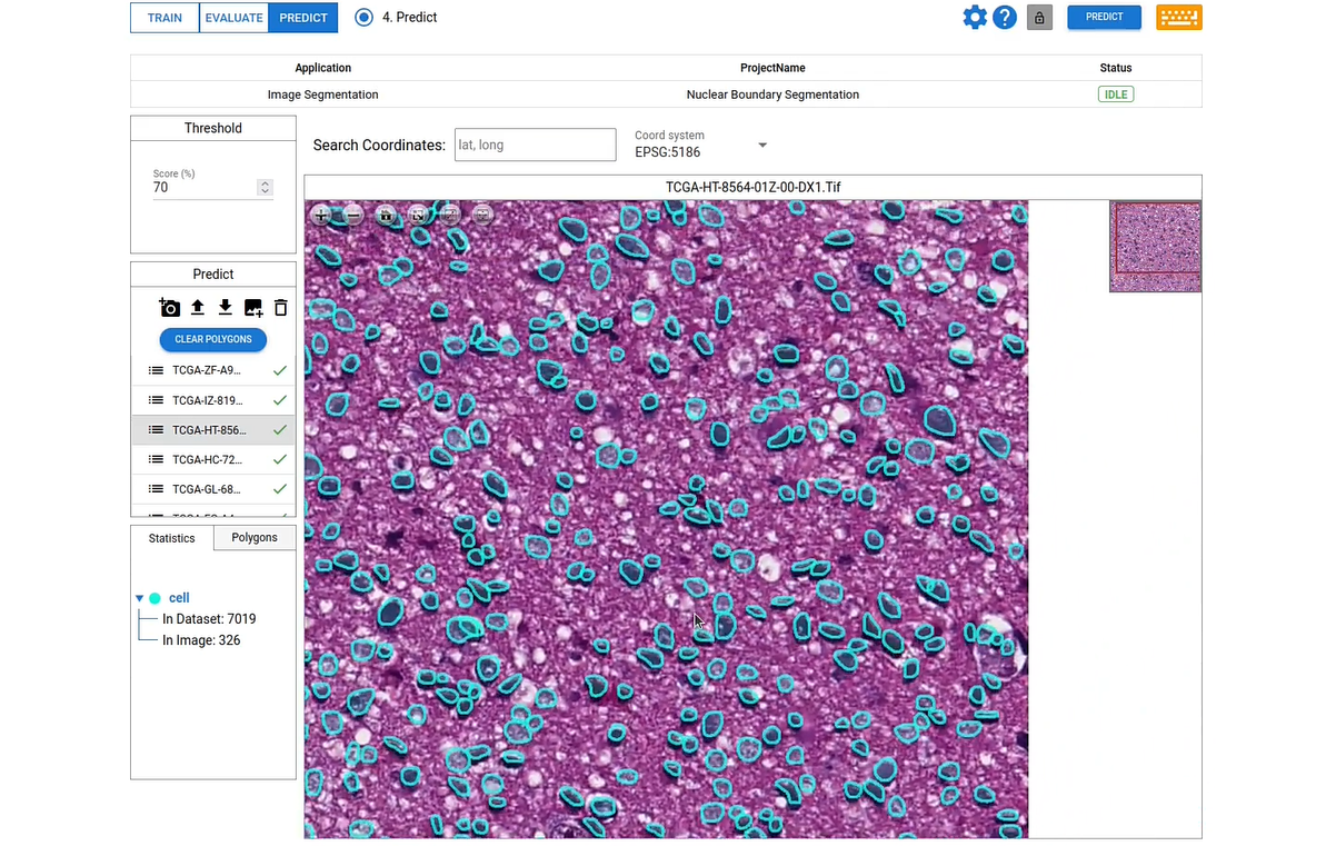
Accurate segmentation of cell nuclei plays a pivotal role in the analysis of microscopic images for medical research. It involves separating the individual nuclei from the background and other cellular components. This segmentation is essential for various applications in biomedical research, including cell counting, cell tracking, and cancer diagnosis. By accurately identifying and outlining the boundaries of cell nuclei, researchers can extract meaningful quantitative data and gain insights into cellular behavior.
Check our latest machine learning project for cell nuclei segmentation from our website.
https://app.deepblock.net/deepblock/store/project/plilk8byvlq6lyc0h
Purpose and Application of Cell Nuclei Segmentation
The purpose of cell nuclei segmentation is to extract relevant information from microscopic images. By accurately segmenting cell nuclei, researchers can perform various analyses such as cell counting, measuring cell size and shape(cell nuclei morphology), and studying cellular dynamics. This information is valuable for understanding cellular processes, identifying abnormal cell behavior, and aiding in the diagnosis and treatment of diseases such as cancer.
Cell nuclei segmentation finds applications in various fields, including biomedical research, drug discovery, pathology, and cytology. In cancer research, for example, accurate segmentation of cell nuclei can help identify cancerous cells, determine tumor grade, and monitor treatment response. In drug discovery, cell nuclei segmentation can be used to assess the effects of potential drugs on cellular behavior.
Importance of Studying Cell Nuclei Morphology
Studying cell nuclei morphology is of paramount importance in various fields of biology and medicine due to the crucial role the cell nucleus plays in cellular function and health. The morphology, or structural characteristics, of cell nuclei provides valuable insights into different aspects of cell biology. Here are some key reasons why studying cell nuclei morphology is important:
-
Cellular Function and Regulation:
- The cell nucleus houses genetic material, including DNA, which contains the instructions for cellular processes and functions.
- Nuclei morphology reflects the state of a cell, its activity, and its potential for functions such as gene expression and cell division.
-
Disease Diagnosis and Prognosis:
- Abnormalities in nuclei morphology are often associated with various diseases, including cancer. Cancer cells often exhibit altered nuclear size, shape, and structure.
- Pathologists use nuclear morphology to diagnose and classify diseases, assess the stage of cancer, and predict patient outcomes.
-
Cell Cycle and Division:
- Changes in nuclear morphology are indicative of different stages of the cell cycle. Studying these changes helps in understanding the regulation of cell division.
- Aberrations in nuclear morphology can signal problems in cell cycle control, potentially leading to uncontrolled cell growth (cancer).
-
Aging and Senescence:
- Cellular senescence, a state of irreversible growth arrest, is associated with changes in nuclear morphology. Studying nuclei morphology can provide insights into the aging process and age-related diseases.
-
Developmental Biology:
- During embryonic development, changes in nuclei morphology are critical for tissue differentiation and organ formation.
- Understanding how nuclei morphology evolves during development contributes to knowledge about normal growth and differentiation processes.
-
Cellular Stress and Damage:
- Exposure to environmental stressors or damage to cellular structures can lead to changes in nuclear morphology.
- Studying nuclei can help identify the impact of stressors and understand cellular responses to external factors.
-
Drug Development and Toxicology:
- Pharmaceuticals and toxic substances can influence nuclear morphology. Assessing these changes is essential in drug development and toxicology studies.
- Researchers use nuclei morphology as a marker for drug efficacy and potential toxic effects on cells.
-
Genetic and Epigenetic Regulation:
- Nuclear morphology is linked to genetic and epigenetic regulation. Understanding nuclear structure aids in unraveling the complex interplay between genes and their regulation.
-
Neuroscience:
- In neuroscience, studying nuclei morphology is crucial for understanding neuronal development, function, and degeneration.
- Nuclear abnormalities can be indicative of neurodegenerative diseases.
-
Biomedical Imaging and Diagnosis:
- Advances in imaging technologies, such as confocal microscopy and high-resolution imaging, enable detailed analysis of nuclei morphology.
- Imaging nuclear morphology is vital for non-invasive diagnosis and monitoring of diseases
The Advantages of Deep Learning in Cell Nuclei Segmentation
Deep learning has revolutionized cell nuclei segmentation by providing highly accurate and efficient segmentation algorithms. Unlike traditional image processing techniques that rely on handcrafted features, deep learning approaches learn directly from the data and can automatically extract relevant features for segmentation. This eliminates the need for manual feature engineering and improves the robustness of the segmentation algorithms.
The advantages of deep learning in cell nuclei segmentation include improved accuracy, faster processing times, and reduced manual effort. Deep learning models can handle diverse cell types, various staining protocols, and different experimental conditions, making them versatile for different research settings.
Introducing DeepBlock.net: A Revolutionary Tool for Cell Nuclei Segmentation
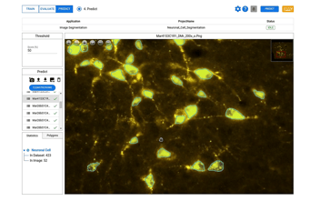
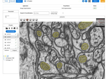
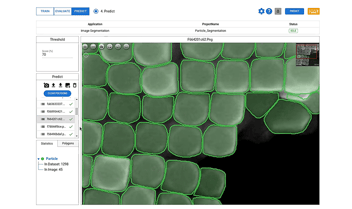
DeepBlock.net is a cutting-edge machine vision software that leverages the power of deep learning for cell nuclei segmentation. It provides researchers with an intuitive and user-friendly interface to perform accurate and efficient segmentation of cell nuclei. DeepBlock.net offers a range of pre-trained deep learning models for life science research, allowing researchers to achieve precise analysis without the need for extensive coding or model training.
With DeepBlock.net, researchers can easily upload their microscopic images, select the appropriate deep learning model, and obtain accurate cell nuclei segmentation results within minutes. Additionally, DeepBlock.net offers annotation tools that enable researchers to train and create their own machine learning models, even without extensive knowledge in computer science. Deep Block no-code AI platform empower researchers to customize and develop their models according to their specific research needs.
Incorporating DeepBlock.net into the cell nuclei segmentation process offers several benefits. It saves time and effort by automating the segmentation task, freeing researchers to focus on higher-level analyses. The high accuracy of the deep learning models ensures reliable results, reducing the risk of manual errors. Additionally, DeepBlock.net is constantly updated with the latest advancements in deep learning, ensuring researchers have access to state-of-the-art segmentation algorithms.


